
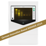


Details

The new way of EXCELLENT gel documentation
FastGene® FAS-X combines:
- modern design,
- state-of-the-art technology using the safe Blue/Green LEDs as light source,
- very user-friendly software,
- high quality Made in Germany.
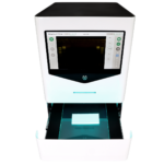
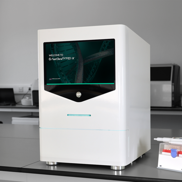
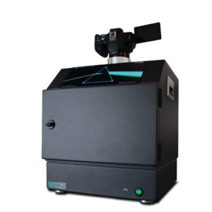
All In One – No additional equipment required
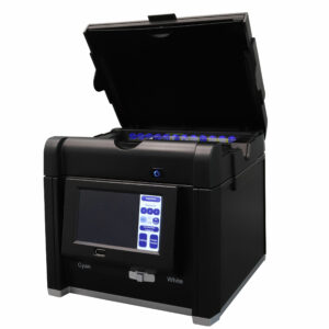
The FastGene® FAS-X is an impressive stand-alone system. Its elegant design and small footprint (only 40 cm) allow it to fit into any laboratory environment. No external computer is required.
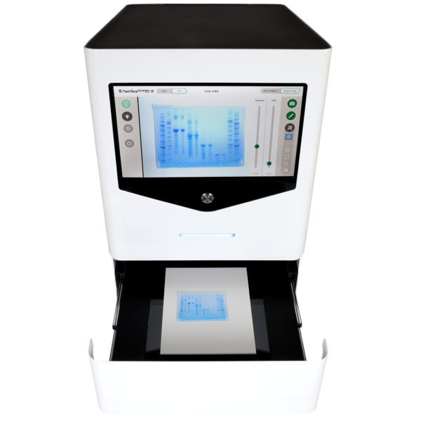
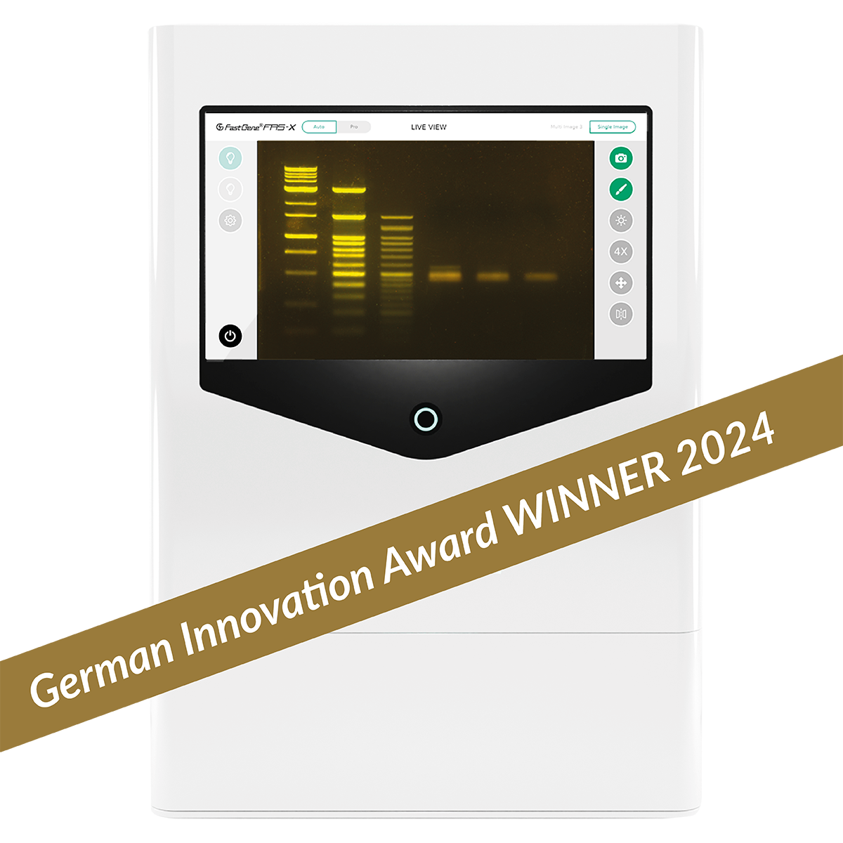
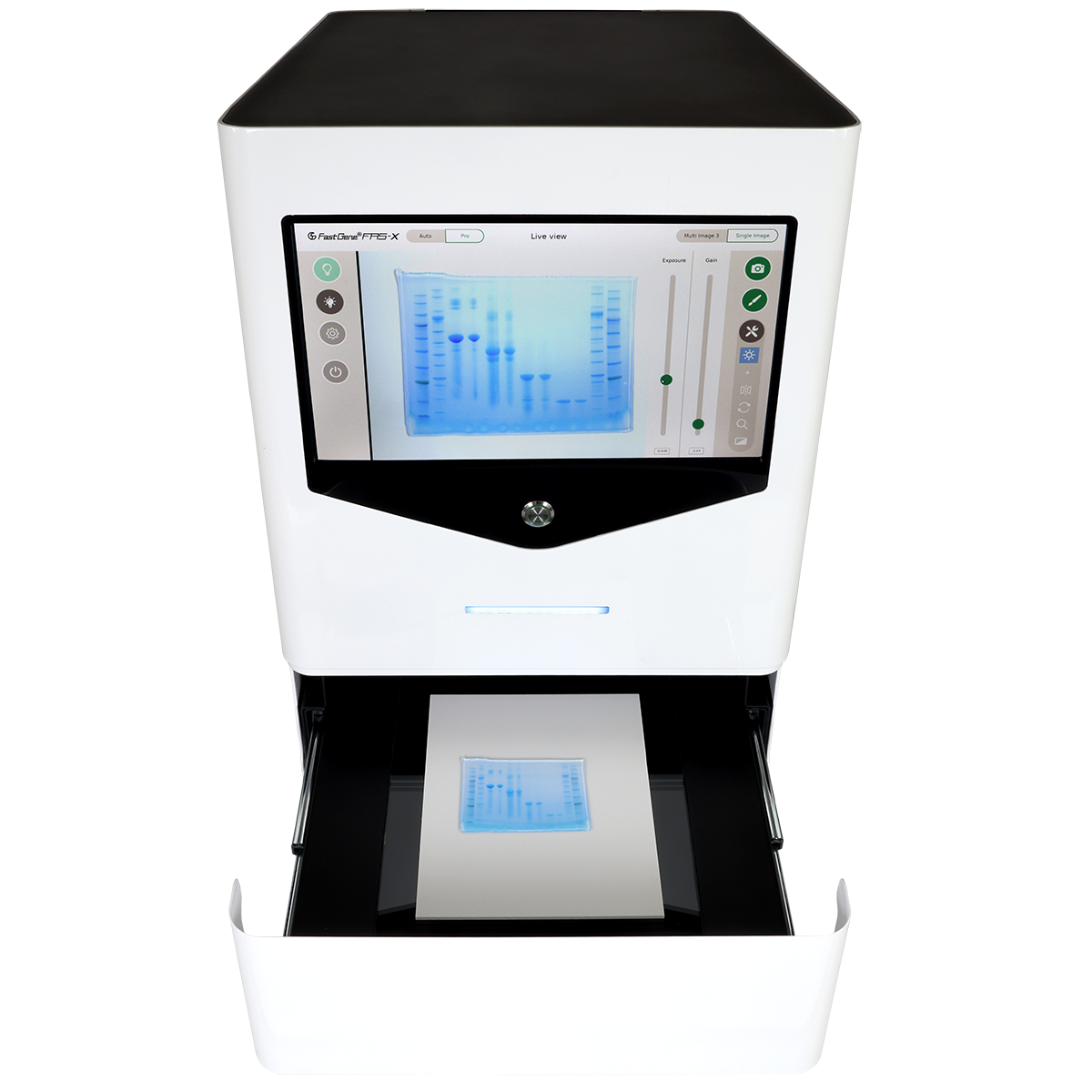
Enhanced interaction via the 13.3” full HD touch screen provides a simple user experience, while large storage capacity (128 GB) and network compatibility improve data management and workflow efficiency.The transilluminator is integrated into a push-to-open loading drawer ensuring absolute darkness and the best image quality without light contamination.
Brilliant image quality
- Equipped with a 20 MPixel colour camera, FastGene® FAS-X provides high resolution gel images and simplifies the process of selecting the area of interest on your gel.
- The camera‘s scientific-grade 1 inch CMOS sensor offers a remarkable sensitivity, detecting nucleic acids at concentrations as low as 2 ng. This ensures that every detail is captured with precision for publication-quality images. Don‘t let any band go unnoticed!
- The giant transilluminator with a viewing area of 26 cm x 21 cm ensures perfect illumination of all gel sizes, large gels as well as multiple gels.
- The prefocused optical unit allows you to capture images without the need to refocus, saving you time and allowing you to capture images faster.
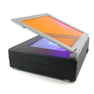
Not UV, not Blue LED, but Blue/Green LED! SAFE gel imaging to protect you and your samples
UV light is dangerous and destructive. Blue LEDs have poor sensitivity to many dyes. With the FAS-X, it is time to say goodbye to these problems! Our unique Blue/Green LED technology protects the user and will boost your results with incredible sensitivity.
- The most important innovation is the implementation of the Blue/Green LED technology (learn more about the technology). No harmful UV-light is used, so there is no risk of UV light exposure!
- Commonly used UV light poses risks not only to the user but also to the sample. Blue/Green LED technology works within the wavelengths of a light spectrum ranging from 470 nm to 520 nm. This range of light is in the visible spectrum and is not harmful to the user or the DNA sample, making the gel documentation process safer.
- One light source for DNA, RNA and proteins! In addition to its remarkable safety, the integrated Blue/Green LED light effectively excites a wide range of common red (incl. EtBr) and green DNA dyes as well as fluorescent proteins with very high intensity. This is achieved by accumulating the light energy absorbed by the fluorophores (figure 1).

Figure 1: BLUE/GREEN LED LIGHT IMAGES: DNA gels stained with green (MIDORIGreen Xtra DNA stain) and red (GelRed® DNA stain) DNA/RNA dyes and a protein gel stained with fluorescent protein dye (SERVA Lighting Red protein stain).
Powerful white light
- Documentation beyond fluorescent gels is possible with the additional white light. The filter wheel in the FAS-X offers the advantage of effortlessly switching between fluorescence (Blue/Green LED light) and the white light mode.

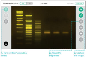
Figure 2: Lighting mode is easily changed using the touch screen buttons.
- Together with the included white plate (figure 3), the white light mode can be used to visualize colourimetric PAGE protein gels with an impressive quality. In addition, high-resolution images of (colourimetric) western blot membranes or bacterial colony plates can be obtained in white light mode (figure 4).

Figure 3: FastGene® FAS-X with open drawer. Coomassie stained PAGE protein gel is placed on the white plate.
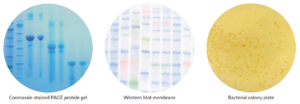
Figure 4: WHITE LIGHT IMAGES: a Coomassie stained PAGE protein gel, a colourimetric Western blot membrane and a bacterial colony plate.
Software makes it easy
- The software has been designed to make operating the instrument and communicating with the camera as easy as possible.
- The user-friendly interface is integrated into the touch screen and allows you to capture the best gel formations with a few simple clicks.
- The software also includes a powerful editing function.
For more detailed information, please refer to the ‘Intuitive software‘ tab on this page.
Password-protected data security
In the constantly changing world of lab tech, safety and data security are top priorities. Equipped with multiple user login and robust password protection, the FAS-X empowers researchers to safeguard their work like never before!
Easy cutting of gel bands
The additionally available Amber Board for FAS-X (FAS-DGOF3) can be easily added to the drawer of the FAS-X to allow safe and convenient cutting of bands.


Dario Lachi, Research Group for Insect Production and Processing, KUL, Leuven, Belgium –
We are currently using the FAS X for several tasks including taking images of DNA gels, protein gels but also all kind of other various things or objects that are part of our studies. We are very pleased with the quality of the images and tend to use the device a lot!
Verified User
Dr. Jeong, S University College of Pharmacy, South Korea –
Our research lab is currently culturing Human Cancer cells, extracting DNA, and using Nippon Genetics Europe FAS-X Gel Doc to confirm the separated bands through Gel using Amplicon.
The biggest reason for choosing this product was to resolve the DNA damage problem that occurred when using existing UV Transilluminators and to obtain desired experimental results.
FAS-X Gel Doc uses UV instead of Blue/Green LED, minimizing DNA damage while providing clear images.
After actually using it, the band images appeared very clear and sharp, and even finely separated bands could be accurately confirmed, which increased the reliability of the experimental results.
When I first considered the size of FAS-X Gel Doc, I thought it was rather large, but after actually installing it in the lab, it occupied less space than other Gel Doc systems, allowing for efficient space utilization.
Also, the intuitive usability based on the Android system greatly improved user convenience, and I was able to easily obtain the desired images without complicated settings.
However, I believe that improvement is needed in the fact that the Android-based system’s stability sometimes decreases, requiring re-booting.
Apart from this point, I am confident that FAS-X Gel Doc is a very satisfactory solution for researchers who want to obtain high-quality band images without DNA damage.
Verified User